Our Posters at UCLA Science Poster Day 2014
The Elegant Mind Club presented a total of 9 posters this year at
- UCLA Science Poster Day 2014
- on May 13, 2014 at Ackerman Grand Ballroom
- during UCLA Undergraduate Research Week, May 12-16, 2014


Dean's Prize
Both Peter and Tim have been awarded the Dean's Prize. Congratulations!


Session 1: 12:00 - 1:00 pm
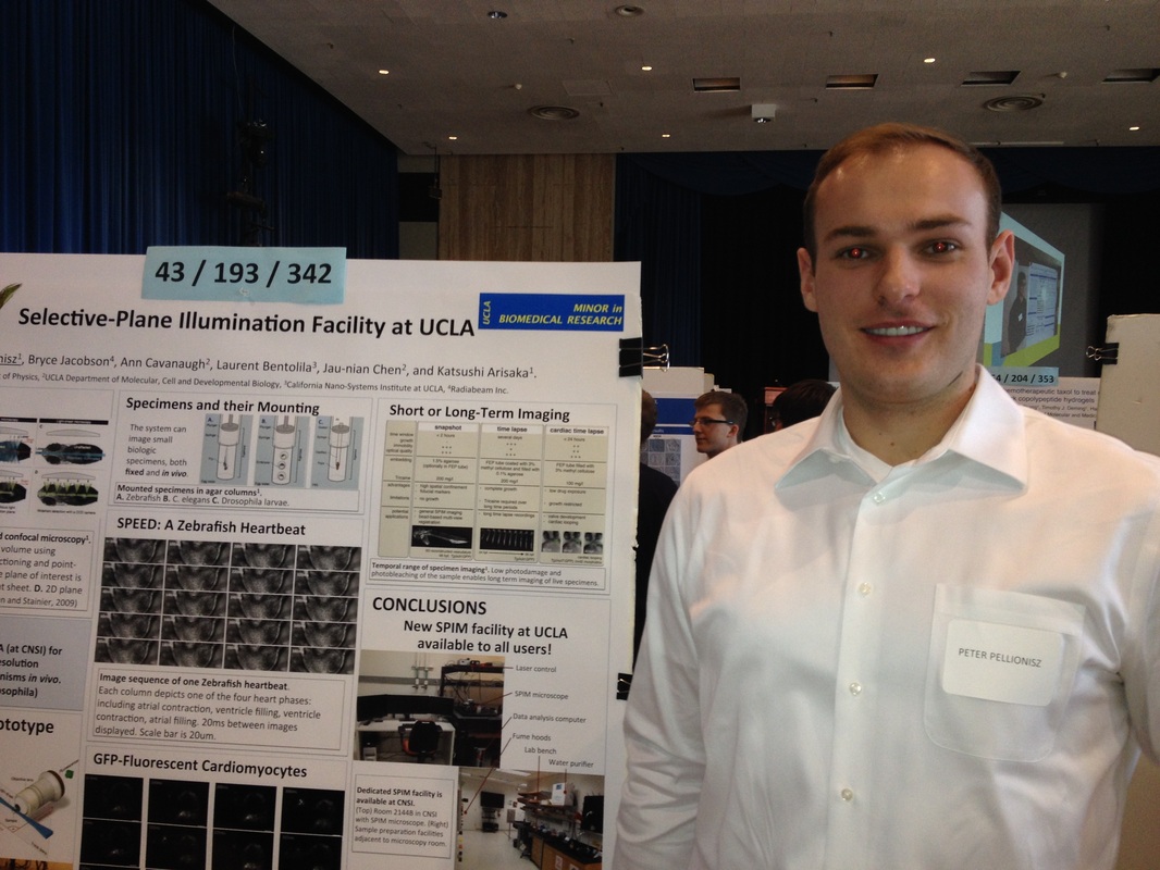
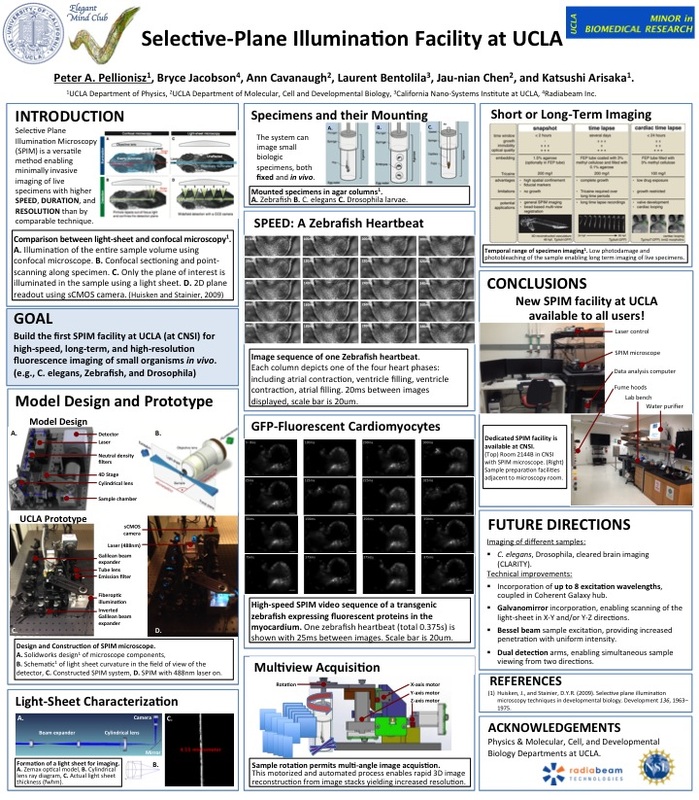
[#41] Selective Plane Illumination Microscopy Enables Visualization of Calcium Ion Flux in the Zebrafish Heart
PETER PELLIONISZ, Bryce Jacobson, Ann Cavanaugh, Laurent Bentolila, Jau-nian Chen, and Katsushi Arisaka.
Defects in calcium efflux and reuptake in cardiomyocytes are associated with heart failure and cardiac arrhythmias. In Zebrafish tremblor mutants, uncoordinated irregular cardiac contractions are due to a calcium extrusion defect. The synthetic compound efsevin is reported to restore persistent, coordinated, and rhythmic contractions in tremblor mutant cardiomyocytes. In order to study calcium gradients in mutant Zebrafish hearts and investigate the role of efsevin regarding calcium handling, a selective plane illumination microscope (SPIM) is constructed. The SPIM system utilizes a light-sheet based orthogonal objective arrangement to provide optical sectioning while reducing phototoxic damage and photobleaching in comparison to conventional confocal techniques. Furthermore, use of a scientific complementary metal-oxide detector (sCMOS) permits high frame rate image acquisition at native resolution. The SPIM system is utilized with fluorescent calcium indicators in genetically engineered Zebrafish to investigate the clearing of calcium from cardiomyocytes following muscle contraction. A significant application of this technique is the fast, high resolution imaging of both live and large-volume biological samples.

| peter-openspipm.pdf |
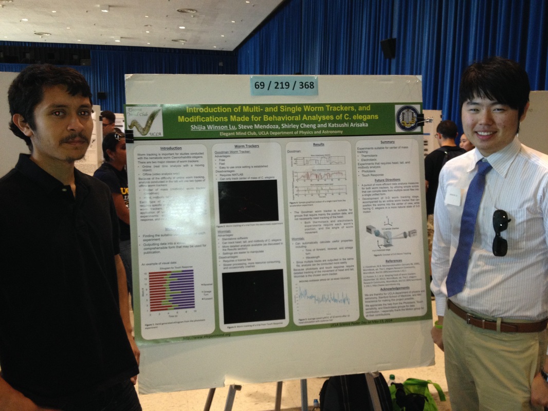
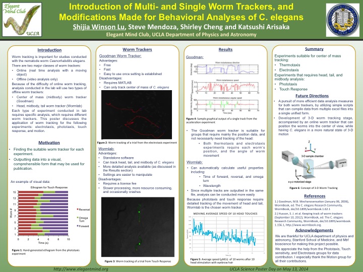
[#69] Introduction of Multi- and Single Worm Trackers, and Modifications Made for Behavioral Analyses of C. elegans
SHIJIA Winson. LU, Shirley Cheng, Steve Mendoza, Katsushi Arisaka
C. elegans is a convenient animal to use in behavioral experiments. In order to characterize behavior, it is necessary to track the motion of these worms over time. We describe a worm tracker that we have used for our experiment to show speed, velocity, and angular position. The worm tracker we show first is a multi-worm tracker, calculating the center of mass for each worm. However, there is a need for higher resolution studies of subtle motions, such as head movement or bending, which may be relevant for the behavioral study of C. elegans. As well as exploring higher definition worm trackers, we have worked in developing single worm trackers. We will describe our progress in modifying our worm tracker for experiments such as detailing the response curve when C. elegans reverses from a harmful stimulus (such as ultraviolet light or touch). We hope that the general worm trackers can further help in the behavioral analysis of C. elegans.
| winson-worm_tracker.pdf |
Session 2: 1:15 - 2:15 pm
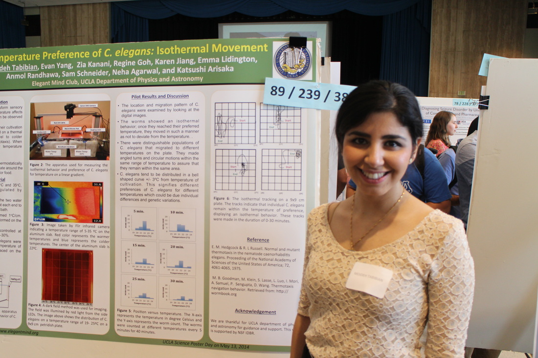
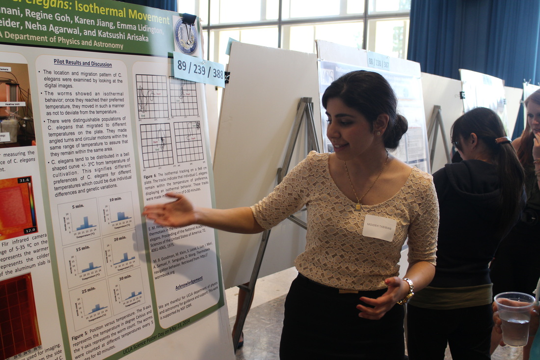
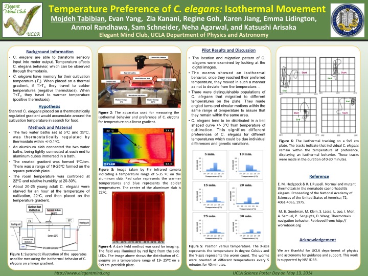
[#239] Preference of Caenorhabditis Elegans for Isothermal Movement
MOJDEH TABIBIAN, Evan Yang, and Katsushi Arisaka
Temperature affects nematode Caenorhabditis elegans navigation behavior, which can be observed through thermotaxis. We examined C. elegans' development of memory for the temperature of cultivation by placing them on a thermal gradient and observing their movements. We expected that starved C. elegans placed on a thermostatically regulated gradient would accumulate around the cultivation temperature in search for food. When we analyzed the movement of C. elegans by taking images, we found that the majority of them were centered on the temperature of cultivation as expected. The C. elegans left a trend traveling along the same temperature, showing preference for that particular temperature. Contrary to our expectation, a few populations of C. elegans were found to prefer accumulating at temperatures other than temperature of cultivation. This variety in navigation behavior may be an indication of individual differences due to genetic activity.
| mojdeh-thermotaxis.pdf |
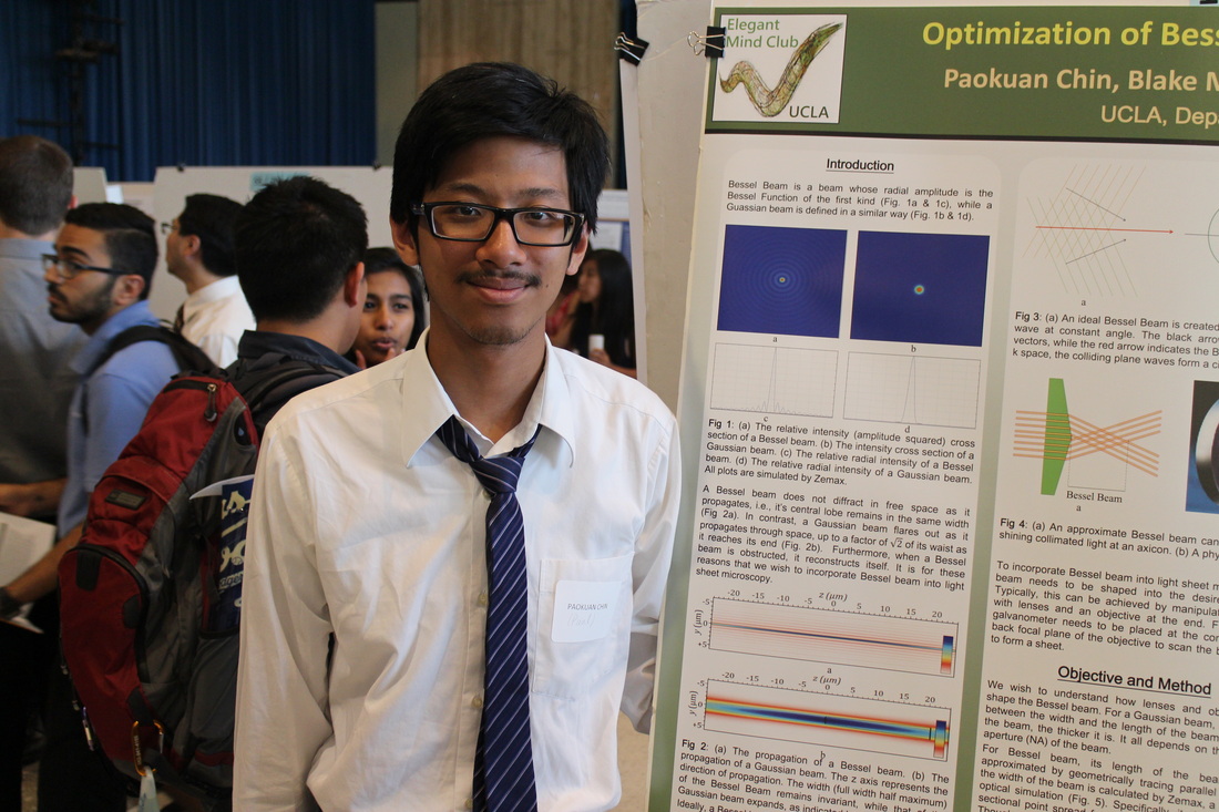
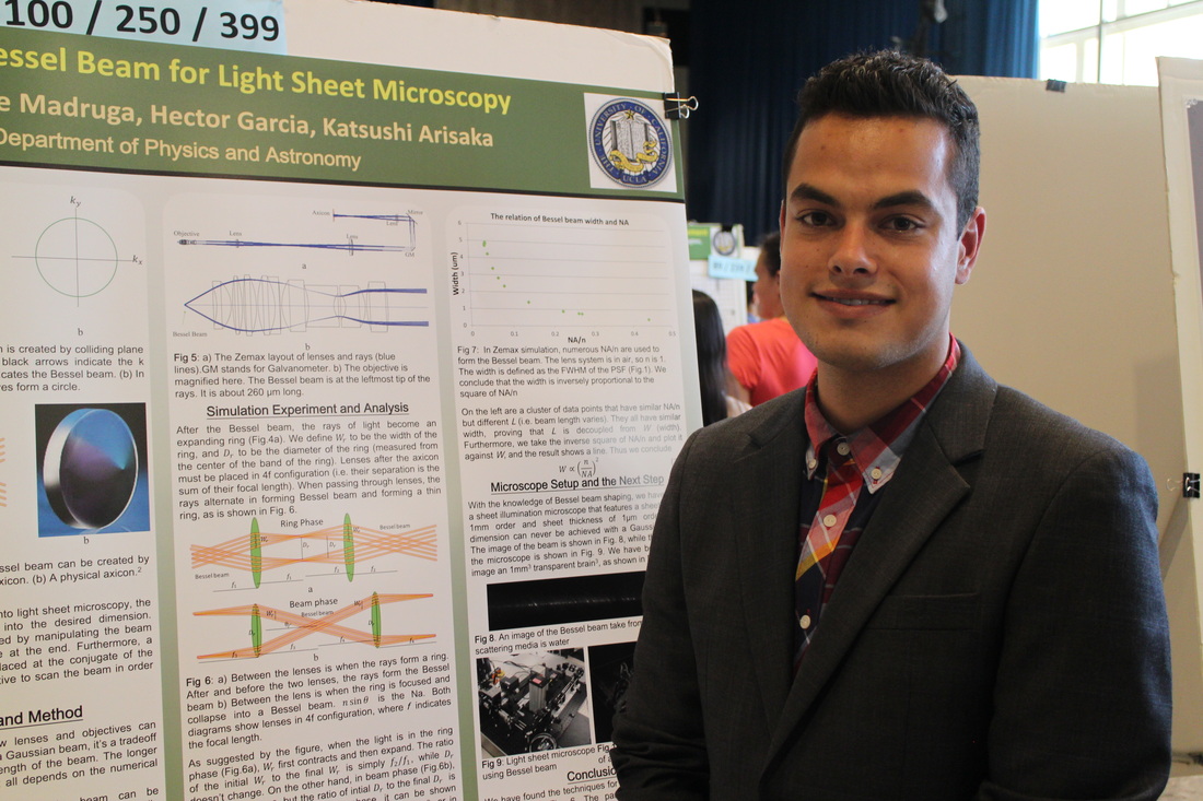
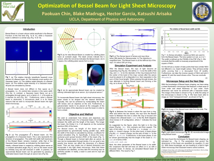
[#250] Optimization of Bessel Beam for Sheet Illumination Microscopy
PAOKUAN CHIN, Blake Madruga, Katsushi Arisaka
Bessel Beam is praised as the self-reconstructing beam. Its virtue lies in its invariant thickness along the propagation axis and the ability to reconstruct itself when obstructed. Sheet microscopy can utilize Bessel beam by scanning it across a plane to create a light volume of remarkable surface area and minimal volume. This advantage results in an especially thin sheet, in order to optically slice samples in question along the previously defined plane. A Bessel Beam can be created by an axicon, a cone shaped lens, or by an annular slit. The former one allows much higher photon efficiency. In the latter method, two parameters that govern the length and thickness of the Bessel Beam have already been mathematically calculated. But that is not the case for Bessel Beam constructed by the former method. In this paper, we report the two parameters of that case. We found them empirically by simulation in Zemax, a program for optical design. With these two parameters in hand, one can shape a Bessel Beam specific for the need of imaging certain samples. However, the Bessel Beam is often condemned for the high percentage of photons residing in its surrounding rings. We verify this property and propose two solutions to this issue.
| paul-bessel_beam.pdf |
Session 3: 2:30 - 3:40 pm
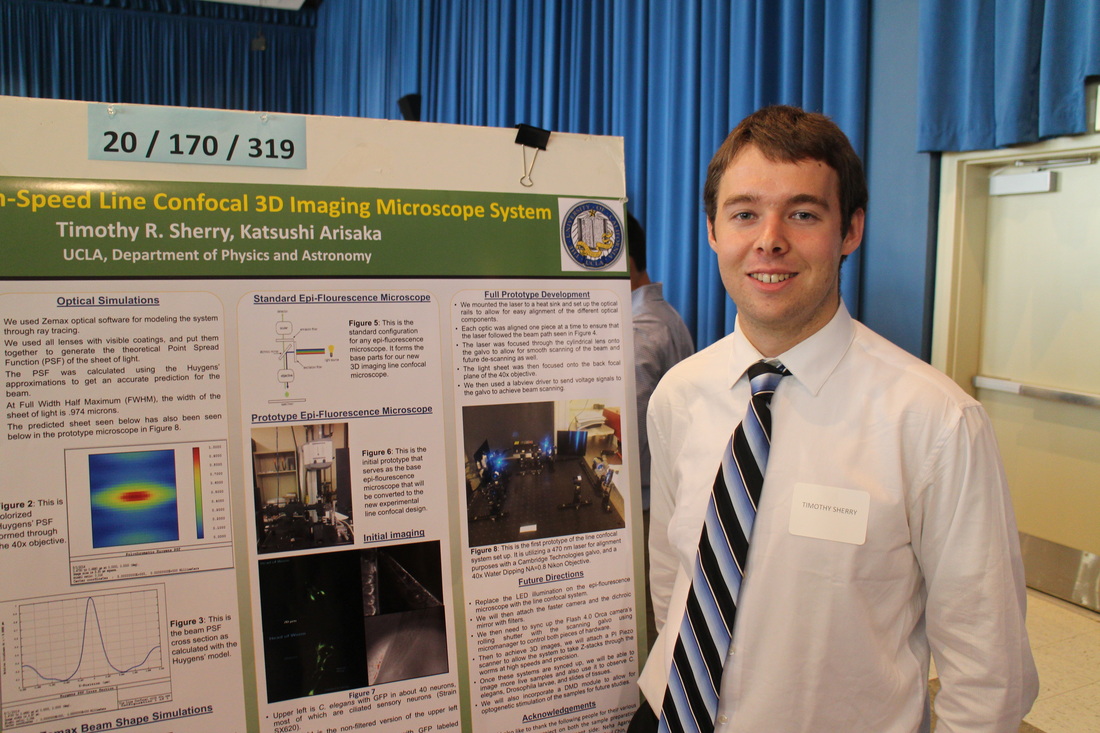
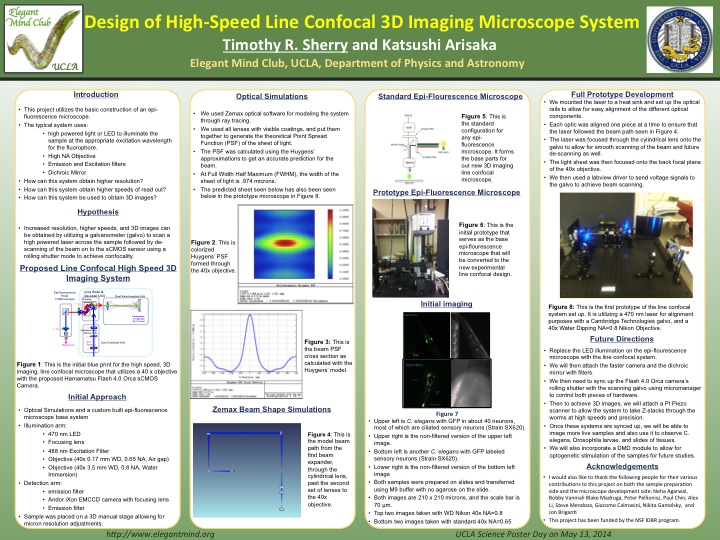
[#319] Design of Line Confocal Microscope High-Speed 3D Imaging
TIMOTHY SHERRY and Katsushi Arisaka
This project seeks to utilize a scientific CMOS camera and sheet illumination to try to capture a 3D image at high speeds relative to other confocal microscope systems. I have utilized a cylindrical lens to create a narrow sheet alongside a galvanometer to scan the beam across the sample quickly to generate the 3D image on the sCMOS sensor. By using the newly developed rolling shutter mode on the sCMOS camera that goes line by line, I am able to achieve confocality without the normal use of a slit. The system utilizes a 470-nanometer laser that can be swapped with a 488-nanometer laser to excite green fluorescent protein to allow neural network imaging of C. Elegans. This system can also be attached to current wide-field microscopes to convert them into line confocal systems allowing for higher resolution at much cheaper prices than a regular confocal microscope as well. The system has performed well in trials, and should present itself as a viable and economical alternative to current confocal microscope systems.
| tim-conforcal.pdf |
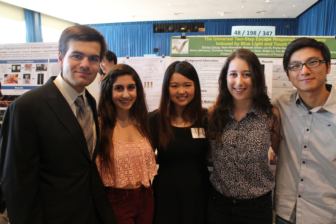
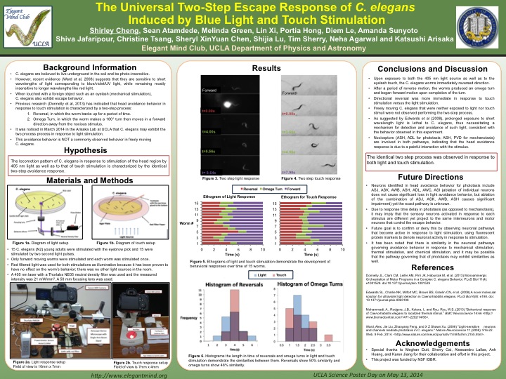
[#347] Similarities of Phototaxis and Mechanotaxis Escape Response in C. Elegans
SHIRLEY CHENG, Shijia Lu, Timothy Sherry, Katsushi Arisaka
Many organisms induce nociception to avoid danger and stressful conditions. A unique and complex nociceptive response has been characterized in C. elegans when stimulated with mechanical and thermal stimuli. More recently, it has also been found that blue and UV light provoked a similar negative phototactic response. However, the connection between light response and nociceptive behavior in C. elegans is unknown. The behavioral response of C. elegans to blue laser light was observed first. Using an AmScope-camera and laser setup, it was found that stimulation of the nose and anterior part of the body elicited a specific escape response that is also observed in the touch response. Upon stimulation, C. elegans moves backwards and then executes a sharp ventral omega turn, changing its direction of locomotion. Previous research on mechanosensation of C. elegans has mapped out the neural mechanisms of that response, which include nociceptors. Future studies will include using a worm tracker program that can compare the dynamic motion of C. elegans to light and touch stimuli. If the data is consistent between the two responses, it can provide convincing evidence that the neural network involved in the different stimuli are related. This study can further our understanding of how sensory transduction is conserved in C. elegans and how this behavior promotes survival by avoiding threatening stimuli.
| shirley-phototaxis.pdf |
[#387] Acceleration Behavioral Experiments Using C.Elegans
DAVID WANG, Steve Mendoza, Shijia Lu, Katsushi Arisaka
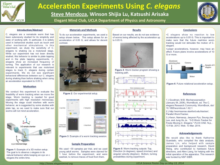
C. elegans are a nematode worm that has been extensively studied for its simplicity and ease of working with. In particular, it is widely used in behavioral studies such as touch and other mechanical stimulations. In this experiment, we study the sensitivity of C. elegans to shaking via a motorized stage. While our experiment has not been directly studied, this behavior is similar to plate tapping and in the plate tapping experiments, C. elegans show an increased frequency of turning. The application of our research is relevant to experiments that use motorized stages to track C. elegans during certain experiments. We do not see significant behavioral differences between our C. elegans during shaking than before shaking when using an acceleration equivalent to 0.05g.
DAVID WANG, Steve Mendoza, Shijia Lu, Katsushi Arisaka
C. elegans are a nematode worm that has been extensively studied for its simplicity and ease of working with. In particular, it is widely used in behavioral studies such as touch and other mechanical stimulations. In this experiment, we study the sensitivity of C. elegans to shaking via a motorized stage. While our experiment has not been directly studied, this behavior is similar to plate tapping and in the plate tapping experiments, C. elegans show an increased frequency of turning. The application of our research is relevant to experiments that use motorized stages to track C. elegans during certain experiments. We do not see significant behavioral differences between our C. elegans during shaking than before shaking when using an acceleration equivalent to 0.05g.
| steve-accerelation.pdf |
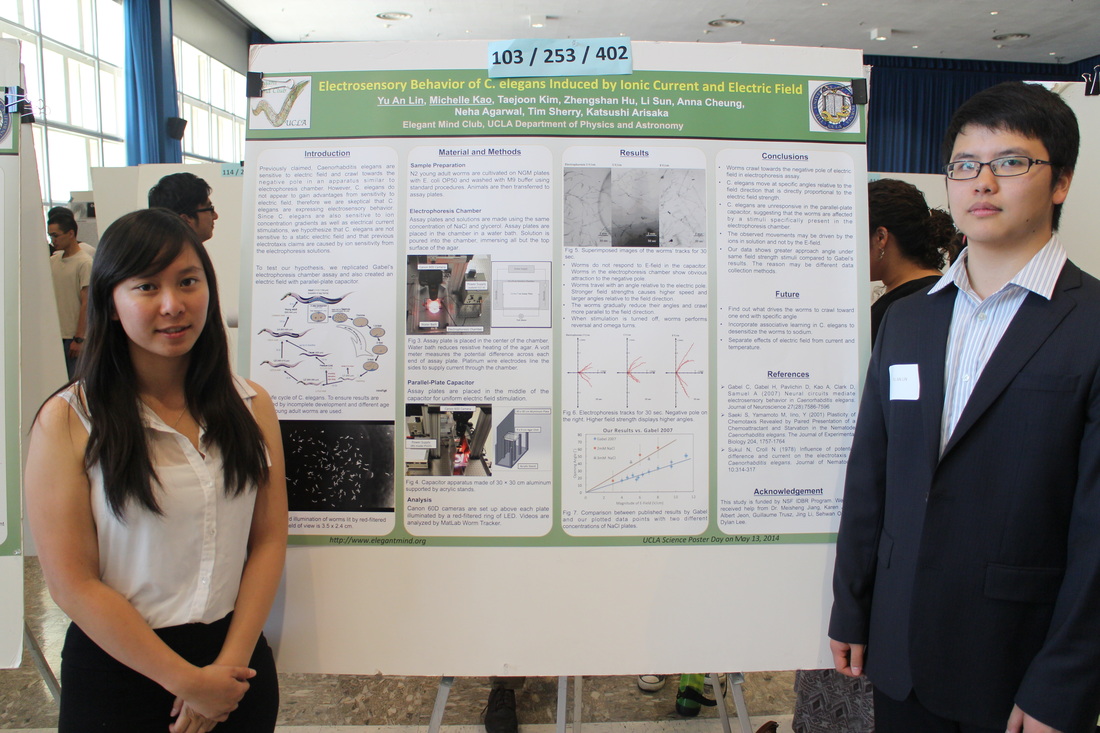
[#402] Quantification of Electric Field Sensory Behavior in Caenorhabditis elegans
YU AN LIN, Michelle Kao, and Katsushi Arisaka
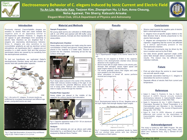
Electric field sensitivity is present in some organisms such as elasmobranchii and monotremes due to their evolutionary advantages. Papers published previously by Gabel et al. claim that Caenorhabditis elegans are also sensitive to electric field and are inclined to crawl towards the negative pole using a set up similar to an electrophoresis chamber. Other studies indicate that C. elegans are sensitive to ion concentration gradients as well as electrical current stimulations. Due to its environment, C. elegans do not appear to gain advantages from sensitivity to electric field, we therefore hypothesize that C. elegans are in fact not sensitive to a pure electric field and that previous electrotaxis claims are caused by ions sensitivity from the electrophoresis solutions. We test this hypothesis by replicating Gabel's experiment and creating our own stimulation with pure electric field using an aluminum parallel plate capacitor. If C. elegans are sensitive to electric field, both stimulations should incite responses in the worms. Using a worm tracking software, we analyzed and quantified the trajectory of individual worms navigating on agar plates of the electrophoresis chamber and compared it to the ones of the parallel plate capacitor. We observed that the worms crawled towards the negative pole in the electrophoresis chamber but showed no response in the capacitor, suggesting that the worms are not sensitive to pure electric field and may be sensitive to the drifting ions in the chambers.
YU AN LIN, Michelle Kao, and Katsushi Arisaka
Electric field sensitivity is present in some organisms such as elasmobranchii and monotremes due to their evolutionary advantages. Papers published previously by Gabel et al. claim that Caenorhabditis elegans are also sensitive to electric field and are inclined to crawl towards the negative pole using a set up similar to an electrophoresis chamber. Other studies indicate that C. elegans are sensitive to ion concentration gradients as well as electrical current stimulations. Due to its environment, C. elegans do not appear to gain advantages from sensitivity to electric field, we therefore hypothesize that C. elegans are in fact not sensitive to a pure electric field and that previous electrotaxis claims are caused by ions sensitivity from the electrophoresis solutions. We test this hypothesis by replicating Gabel's experiment and creating our own stimulation with pure electric field using an aluminum parallel plate capacitor. If C. elegans are sensitive to electric field, both stimulations should incite responses in the worms. Using a worm tracking software, we analyzed and quantified the trajectory of individual worms navigating on agar plates of the electrophoresis chamber and compared it to the ones of the parallel plate capacitor. We observed that the worms crawled towards the negative pole in the electrophoresis chamber but showed no response in the capacitor, suggesting that the worms are not sensitive to pure electric field and may be sensitive to the drifting ions in the chambers.
| leon-electrotaxis.pdf |
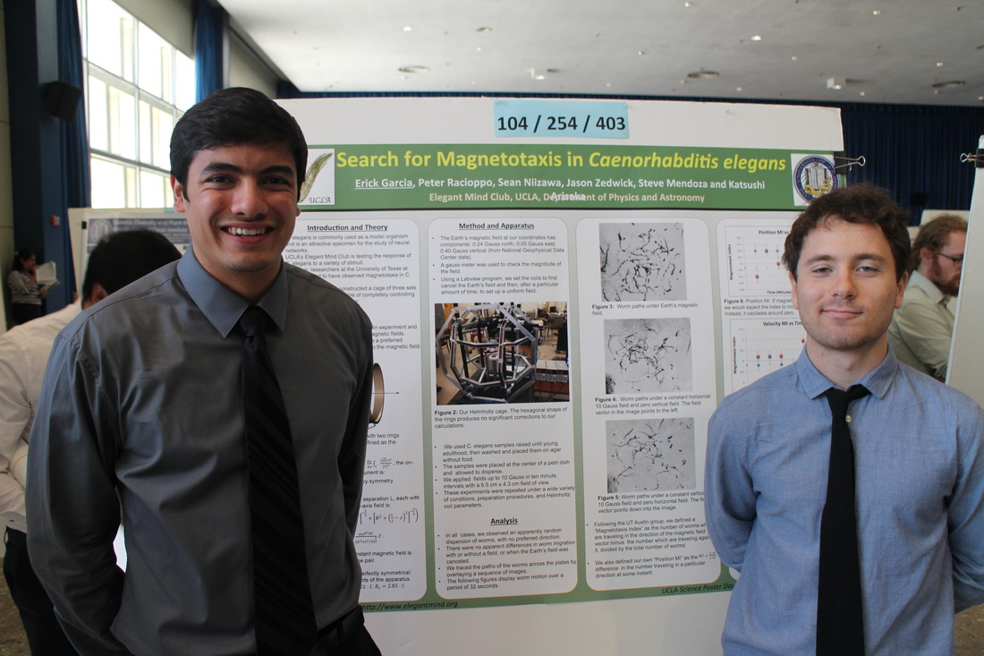
[#403] C. Elegans' Inability to Detect Uniform Magnetic Fields
ERICK GARCIA, Peter Racioppo, Katsushi Arisaka
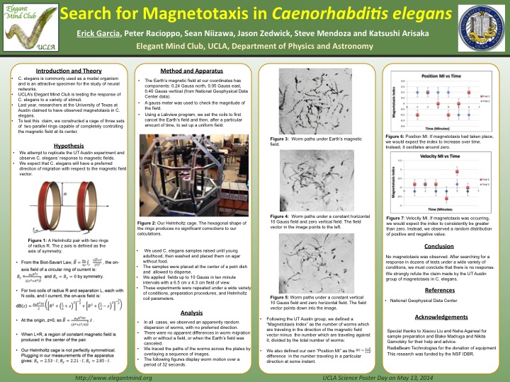
Some organisms have the ability to detect magnetic fields and move as a response to these fields. The nematode Caenorhabditis elegans (C. elegans) was believed to use sensory neurons to detect the earth's magnetic filed in order to orient itself and burrow down within the soil, as opposed to the expected direction of gravity. To test this theory washed, young adult C. elegans were placed in a cage that creates a uniform magnetic field in all three directions of space. They were video recorded under different magnetic field strengths. The cage cancelled the earth's magnetic field for 60 seconds and then a strong magnetic field of five to ten gauss was generated in the same direction as that of the earth for another five to thirty minutes. The strength of the magnetic field generated by the cage was verified by using a gaussmeter, and the direction of the magnetic field was controlled by LabVIEW computer software. C. elegans showed no response to the fields and did not move in the predicted direction or any other constant direction. These results suggest that C. elegans does not possess the sensory abilities to detect uniform magnetic fields, and that another mechanism allows them to orient within the soil.
ERICK GARCIA, Peter Racioppo, Katsushi Arisaka
Some organisms have the ability to detect magnetic fields and move as a response to these fields. The nematode Caenorhabditis elegans (C. elegans) was believed to use sensory neurons to detect the earth's magnetic filed in order to orient itself and burrow down within the soil, as opposed to the expected direction of gravity. To test this theory washed, young adult C. elegans were placed in a cage that creates a uniform magnetic field in all three directions of space. They were video recorded under different magnetic field strengths. The cage cancelled the earth's magnetic field for 60 seconds and then a strong magnetic field of five to ten gauss was generated in the same direction as that of the earth for another five to thirty minutes. The strength of the magnetic field generated by the cage was verified by using a gaussmeter, and the direction of the magnetic field was controlled by LabVIEW computer software. C. elegans showed no response to the fields and did not move in the predicted direction or any other constant direction. These results suggest that C. elegans does not possess the sensory abilities to detect uniform magnetic fields, and that another mechanism allows them to orient within the soil.
| erick-magnetotaxis_rev.pdf |